
What is renal angiomyolipoma (AML)?

A renal angiomyolipoma is a benign (non-cancerous) tumor that forms in the kidneys. It is comprised of three components that are represented in its name. “Angio” refers to the blood vessels that feed the tumor. “Myo” is the smooth muscle and “Lipoma” is the fat that both make up the bulk of the mass.

AMLs can range in size from 2 mm to 30 cm or more in diameter. The smaller varieties often cause no symptoms, but as they grow, they can cause back pain or pain on the sides of the body, blood in the urine (hematuria) and bleeding. Larger AMLs can also cause a noticeable bulge in the abdomen or flank area.
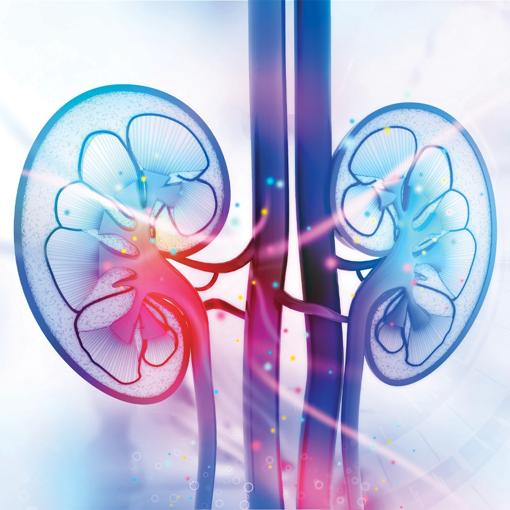

renal angiomyolipoma (AML)

What causes an AML to develop?

AMLs may be caused by mutations in the tuberous sclerosis complex TSC1 or TSC2 genes. These genes act as tumor suppressors, and when they mutate, they can fail to regulate cell growth which results in the development of an abnormal mass. When this occurs, it is typically at a younger age and AMLs develop in both kidneys. AMLs may also be caused by lymphangioleiomyomatosis (LAM), a rare and progressive lung disease.

In some cases, AMLs can develop sporadically, with no family history or underlying genetic conditions or mutations. While rare, AMLs can also occur in people with other genetic conditions such as neurofibromatosis or Hippel-Lindau disease.

Risk factors for AML:

  • Female sex
  • Middle adulthood (between age 30 and 60)
  • Tuberous sclerosis complex (TSC)
  • Lymphangioleiomyomatosis (LAM)
  • Genetic mutations involving the TSC1 or TSC2 genes

How are renal AMLs diagnosed?

Many AMLs are found when an imaging test (such as MRI, CT or ultrasound) is performed for an unrelated reason. This is called an “incidental finding.” If the AML is small and not causing any symptoms, it may be watched over time using diagnostic imaging. Others with underlying genetic conditions may begin monitoring for AMLs using a combination of imaging, blood tests and physical examinations.

Treatment options for renal angiomyolipoma

Many AMLs do not require any immediate treatment because they are small and are not causing any symptoms. Your healthcare provider may recommend “active surveillance” using imaging if the tumor is considered to have a low risk of bleeding.
For those with AMLs related to tuberous sclerosis complex, medications such as mTOR inhibitors may be prescribed to shrink the tumor.

Minimally invasive treatment for AMLs

Renal angiomyolipoma embolization is a minimally invasive treatment performed by the interventional radiologists at University Radiology Vascular & Interventional Center. The procedure blocks the flow of blood to the AML, which shrinks the tumor and can relieve symptoms. This procedure preserves normal kidney function.

Surgery

For some patients, surgery may be recommended. This is typically the approach used when the tumor is large or if less invasive treatments are not successful. If practical, a partial nephrectomy can remove the tumor while preserving the rest of the kidney. In other cases (especially if cancer is strongly suspected), a radical nephrectomy—removal or the entire kidney—may be recommended.

Learn About Your Renal Angiomyolipoma (AML) Treatment Options

For more information on Renal AML embolization, reach out to our New Jersey radiology clinic.